Towards Mixed-State Coded Diffraction Imaging

Benjamin Attal, Matthew O'Toole

Abstract

Coherent diffraction imaging (CDI) is a computational technique for reconstructing a complex-valued optical field from an intensity measurement. The approach is to illuminate an object with a coherent beam of light to form a diffraction pattern, and use a phase retrieval algorithm to reconstruct the object’s complex transmittance from the measurement. However, as the name implies, conventional CDI assumes highly coherent illumination. Recent works therefore extend CDI to account for partial coherence and imperfect detection, by modeling light as an incoherent mixture of multiple fields (e.g., multiple wavelengths) and recovering each field simultaneously. In this work, we make strides towards the practical implementation and usage of multi-wavelength diffraction imaging. In particular, we provide novel analysis of the noise characteristics of multi-wavelength diffraction imaging, and show that it is preferable to coherent diffraction imaging under high signal-independent noise. Additionally, we present a compact coded diffraction imaging system and corresponding phase retrieval algorithms to robustly and simultaneously recover complex fields representing multiple wavelengths. Using a novel mixed-norm color prior, our prototype system reconstructs a larger number of multi-wavelength fields from fewer measurements than existing methods, and supports applications such as micron-scale optical path difference measurement via synthetic wavelength holography.


Multi-wavelength Reconstruction Results through Coded Diffraction Pattern Imaging

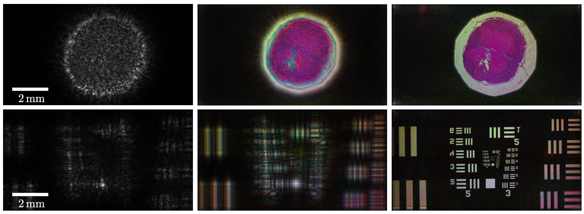
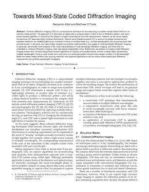
Our proposed imaging technique can recover mutually incoherent modes from a partially coherent field. Below, we demonstrate the ability to simultaneously reconstruct three modes, each at a different wavelength: 638nm (red), 520nm (green), and 445nm (blue). Each individual mode is fully coherent, but because there is no observable interference between the light at these different wavelengths, they are added together on an intensity basis. (Left) We show one of the raw captured images, which is a sum of the intensities of each color at the sensor plane. (Center) We show the amplitude of the reconstructed color modes at the SLM plane. (Right) We digitally refocus the captured field to the object plane. (Top) The first row represents a microscope slide of a rabbit spinal cord (48 measurements), and (Bottom) the second row is of a USAF resolution chart (48 measurements). Note that we do not make use of any filtering optics to reconstruct these images.

Mixed-state CDP

More Details

Please refer to our paper for details. Code and data will be released shortly.

paper

Link to paper


Sponsors

We thank the following for helpful discussions: Wei-Yu Chen, Anat Levin, Aswin Sankaranarayanan, Gordon Wetzstein, and the anonymous reviewers. The authors acknowledge the support of an Uber Fellowship, and NSF Grants IIS-1900821 and IIS-2008464.